Immunofluorescence Microsopy...
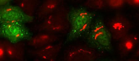
...is a fancy way to say "making my cells glow in the dark." This is what I have to do for the bulk of my current project. I take some cells, make them express my protein (which glows green), label different cellular structures/organelles (which glow red), and try to see where the two colors overlap. This way, I can try and figure out which particular structure/organelle my protein localizes in, which can in turn help me to figure out what its potential role or function might be. So far, I've found nothing too terribly convincing. My protein appears to be EVERYWHERE in the cell (the whole cell just glows neon green), so I'm trying to label it with the green-fluorescent-protein tag (ie GFP tag) in a different way to see if I can try to make my experiments work. I just wanted to share these photos with you cuz I thought that they looked way cool, despite my experiments not working too well. Ali, I know that you are sick and tired of seeing images like this, but you know what? I don't care. It's pretty new and slick to me and I'm guessing that most of you will not have had a chance to see stuff like this.In this first picture, the cells are stained red for the Golgi apparatus. I don't know how much high school bio you remember, but just in case, the Golgi is where newly-synthesized proteins go to be processed and packaged for export. The four cells which are glowing green are expressing my protein but as you can see, it doesn't really co-localize with any of the red that's also stained in those particular cells.
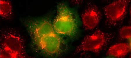
Here, the cells were stained red for ERGIC proteins, which are present in vesicles transporting proteins between the Endoplasmic Reticulum (where new proteins are synthesized) to the Golgi. In the green cells, you can see where there is a tiiiiny bit of overlap between the red and the green (in these cells, the red staining looks kind of orangish rather than red) which could mean that my protein may localize in these transport vesicles. Wheeee! An "almost" result!
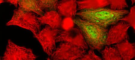
This final pic I just included because it looks really nifty. The red staining corresponds to microtubules, which are present throughout the entire cell and help give it its shape. They make up what is essentially the skeleton of the cell.
On a side note, without warning, my computer has died again and refuses to start up. I am too lazy and too tired of this constant tug-of-war with my piece o' junk HP to try and fix it now. The other blog entries that I had planned require photos which are on my defective computer's hard drive so instead, I took photos off of my lab computer. Your consolation prize? This bio-post.


0 Comments:
Post a Comment
<< Home